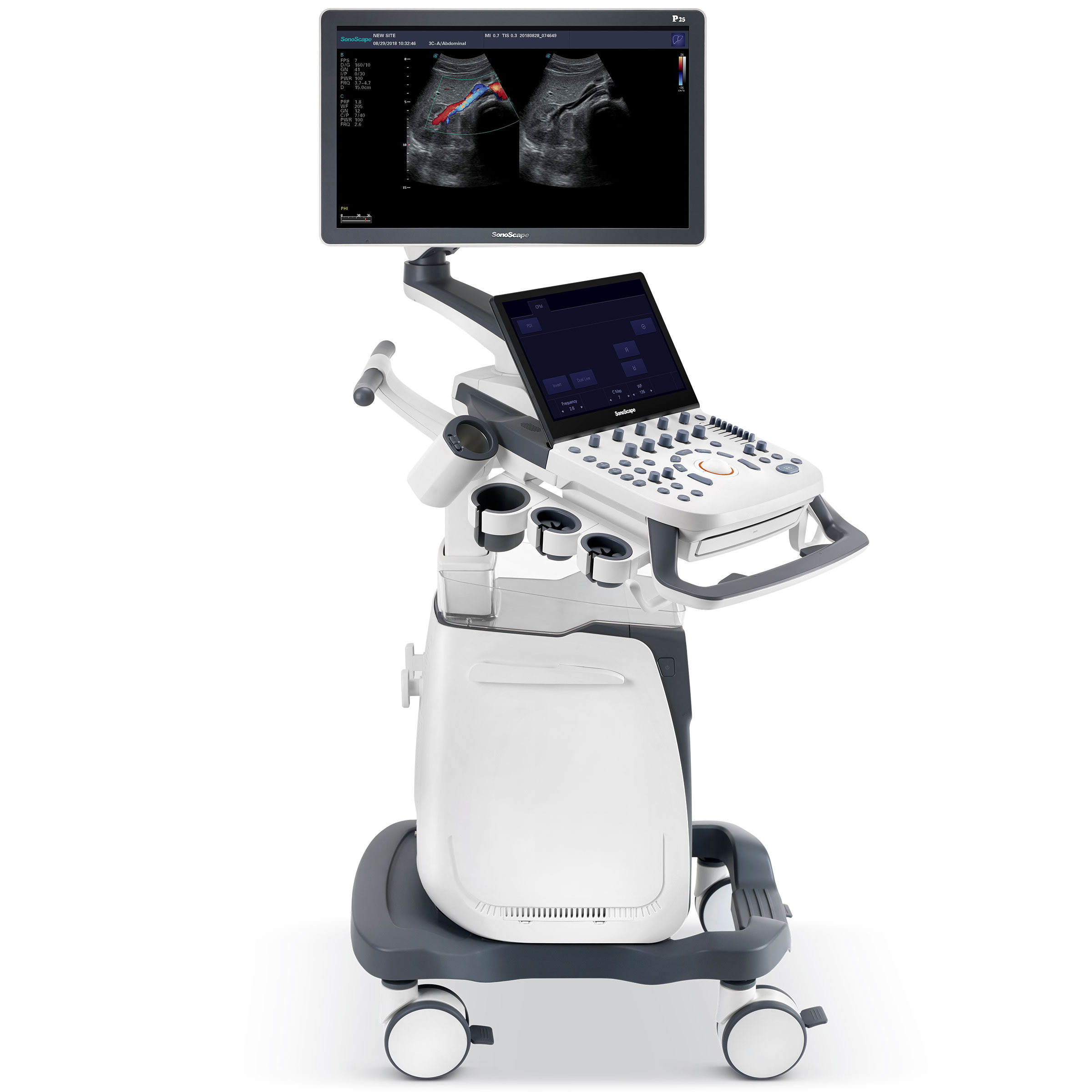

S50 – Updated Range Available
SonoScape’s S50 carries a comprehensive, upgraded platform, to become an epoch-making ultrasound system….equipped with powerful single crystal transducers
- Description
Clarity through improved uniformity and sensitivity
SonoScape’s S50 is equipped with a wide band single crystal probe for abdominal and cardiac scanning, which can greatly improve signal to noise ratio, and acquire stunning images with better resolution and richer imaging detail. Compared with a conventional transducer, a single crystal probe has significantly improved acoustic energy conversion capacity, which means the probe has better performance as well as a longer working life.
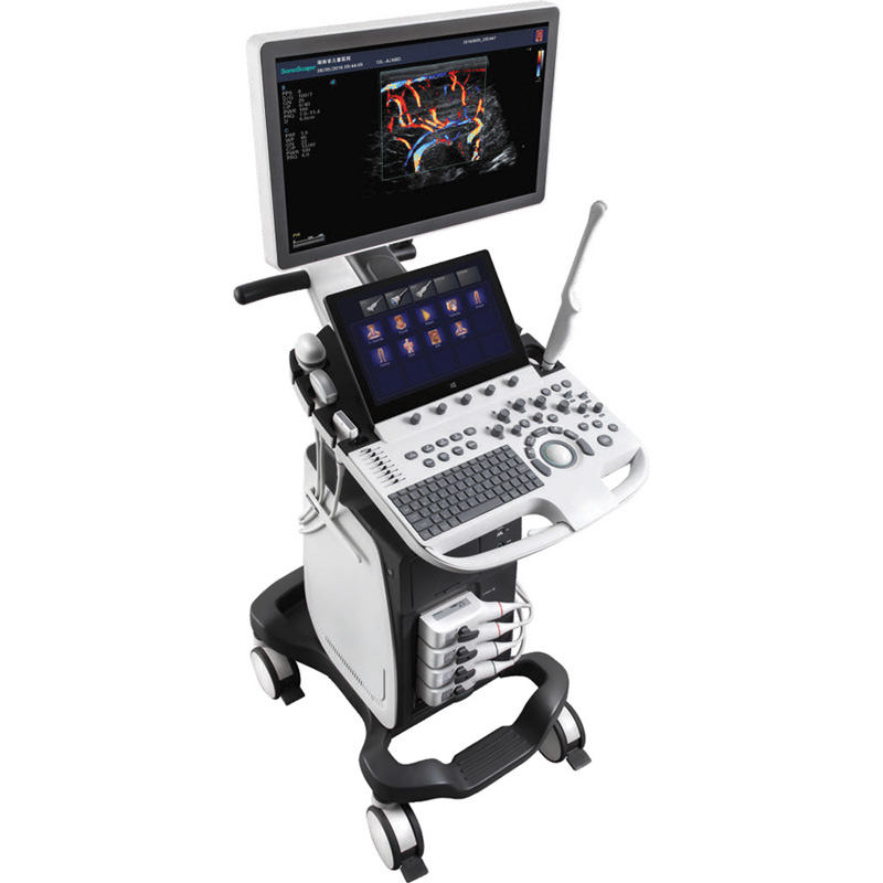
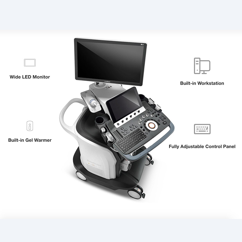
Thoughtfully designed.
Ergonomic design, excellent man-machine interaction and rapid response, makes S50 an intelligent scanning assistant for you, bringing improved efficiency and helping to prevent fatigue from multiple examinations.
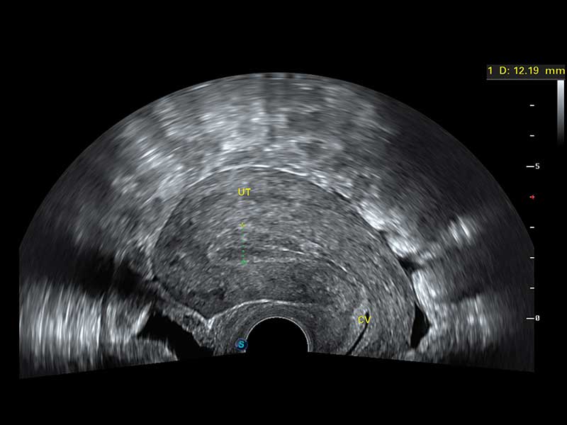
C-xlasto imaging
By understanding that tissue stiffness varies depending on the type of tissue, we can use this to easily find abnormalities and tumors within soft tissue. When applying consistent and repeated pressure with the transducer on soft tissue the system can quickly calculate the Strain Ratio of the region and give an Intelligent Strain Curve for a recording of applied pressure to assist with diagnosis of the abnormality. Predominately used only with linear probes, SonoScape’s new transvaginal probe for gynecological is breaking the mold and expanding elastography applications to non-linear probes.
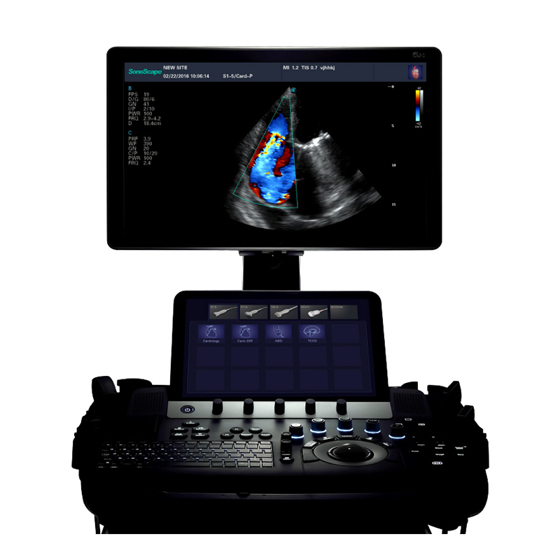
Contrast imaging
Contrast Imaging is the use of contrast agents that are small enough to go into the smallest of veins in the body and still provide a loud signal reflection for the ultrasound to pick up. Such a loud signal reflection allows the ultrasound to accurately display small veins in the body, blood profusion in different organs, and helps to measure blood flow. The S50 takes it a step farther by providing better resolution and deeper penetration, maximizing the short lifespan of the contrast agent bubbles by giving a clearer, more complete image of the desired organ.
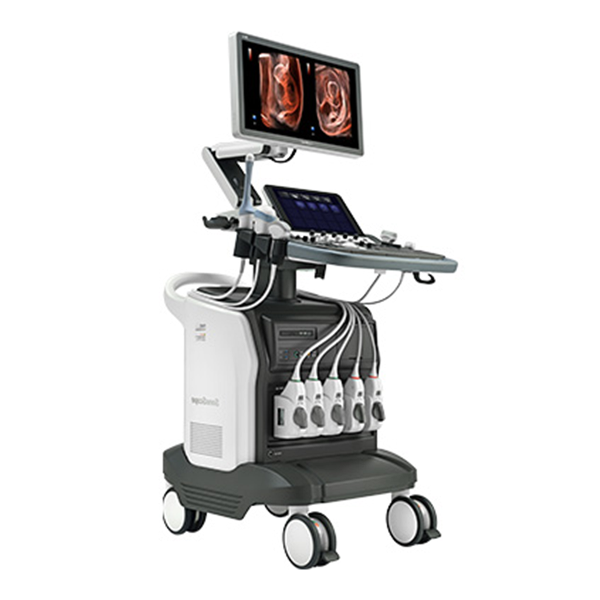
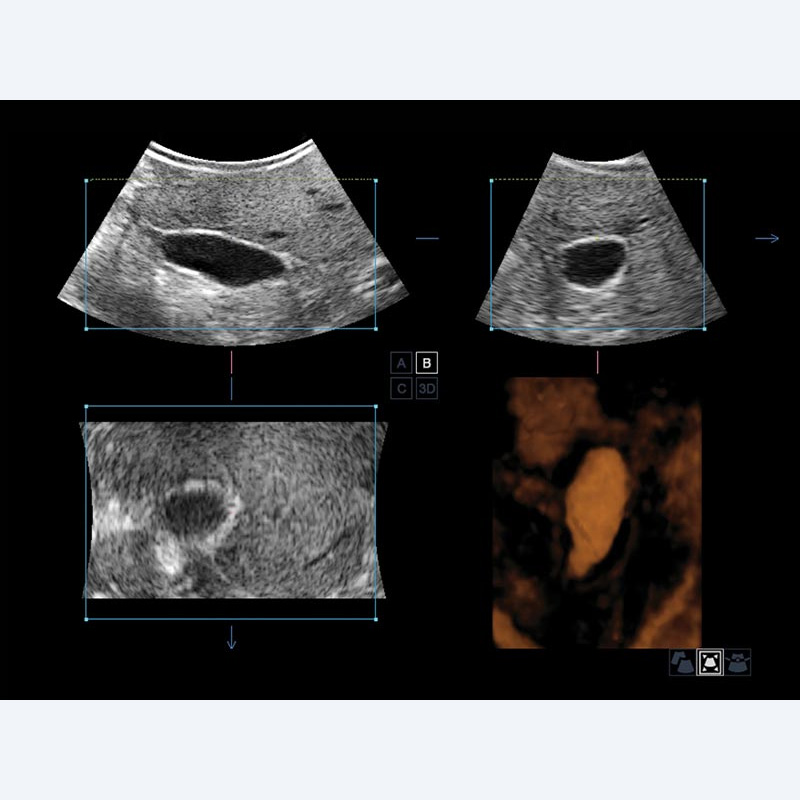
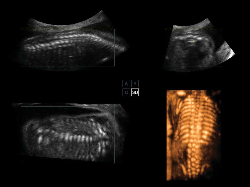
SonoScape’s S50 is configured with a new imaging engine, which can significantly optimize image performance, especially for 3D/4D imaging with speed and convenience. Outstanding volume performance makes S50 outshine others on volume imaging, and dramatically enhances diagnostic confidence.
- Features
Inversion 4D
- It provides more in-depth evaluation of vascular and cystic strictures creating a three-dimensional cast-like volume of the anatomy of interest.
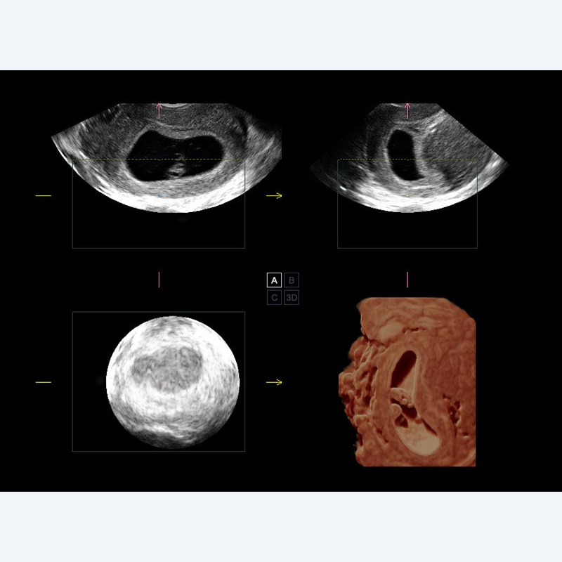
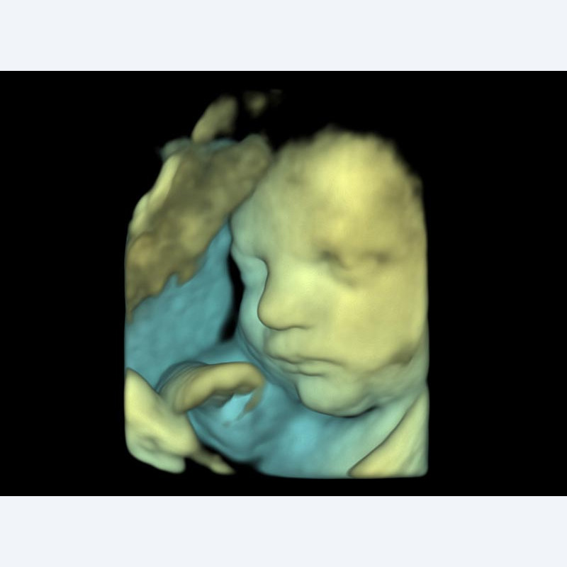
S-Live
- It allows for detailed visualization of subtle anatomical features, thereby enabling intuitive diagnosis on the real-time 3D images and enriching patient communication.
S-Depth
- It can automatically display the near and foe distance relation from transducer to target, presented by smart designed color coding. It can help doctors to judge the spatial relationship on real-time 3D images.
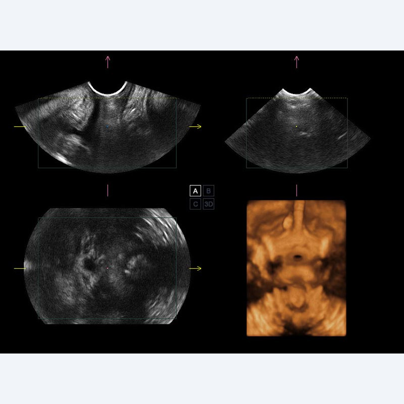
Pelvic Floor 4D
- Transperineal 4D pelvic floor ultrasound can provide useful clinical values in assessing the vaginal delivery impact on the female anterior compartment, judging whether the pelvic organs are prolapsed or not and the extent, determining if the pelvic muscles were torn accurately.
Auto IMT
- Auto IMT is used when determining the level of vascular sclerosis present in the patient by automatically tracing and calculating the thickness of the carotid vessels.
Tissue Doppler Imaging
- Tissue Doppler Imaging allows you to quantitatively evaluate local myocardial movements and functions, with speed and strain/strain rate parameters.
Stress Echo
- Stress echocardiography is the combination of 2D echocardiography with a physical, pharmacological or electrical stress of the patient. It also provides users with report management tools such as configurable template editor, multiple loops to select one for storage, wall motion scoring, stress echo report, etc…